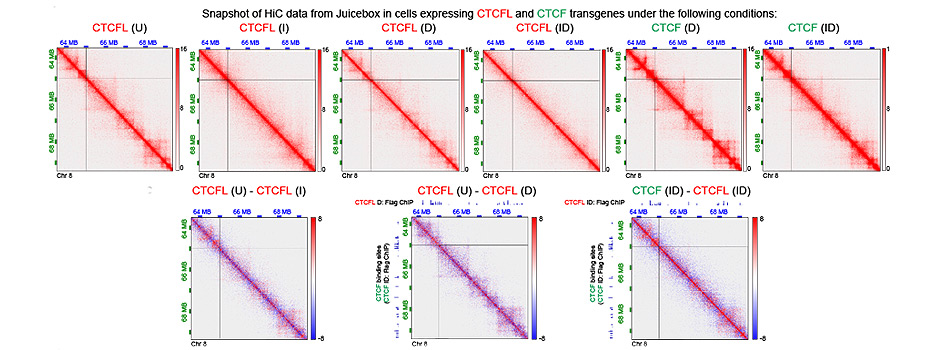
CTCF and CTCFL (BORIS)
We defined where the two proteins overlap and where they differ in binding, insulation and promoter control. This explains when CTCFL can substitute for CTCF, and when it works against it.
We investigate how CTCF, CTCFL, cohesin, condensin II and histone H1 variants shape genome folding, nucleosome organization and gene regulation. We ask how cells buffer transcription against architectural disruption, and how these mechanisms influence development, cancer and developmental disorders. In HER2-positive breast cancer, we use single-cell multiome analyses and computational modelling to investigate how drug combinations prevent the emergence of drug-tolerant persister (DTP) cells.
To uncover how genome organization and chromatin regulate and buffer gene expression, how these mechanisms support development, and how their disruption contributes to cancer and developmental disorders. We investigate CTCF, CTCFL, cohesin, condensin II and histone H1 variants, connecting their roles in genome folding and nucleosome organization to transcription and cell identity.
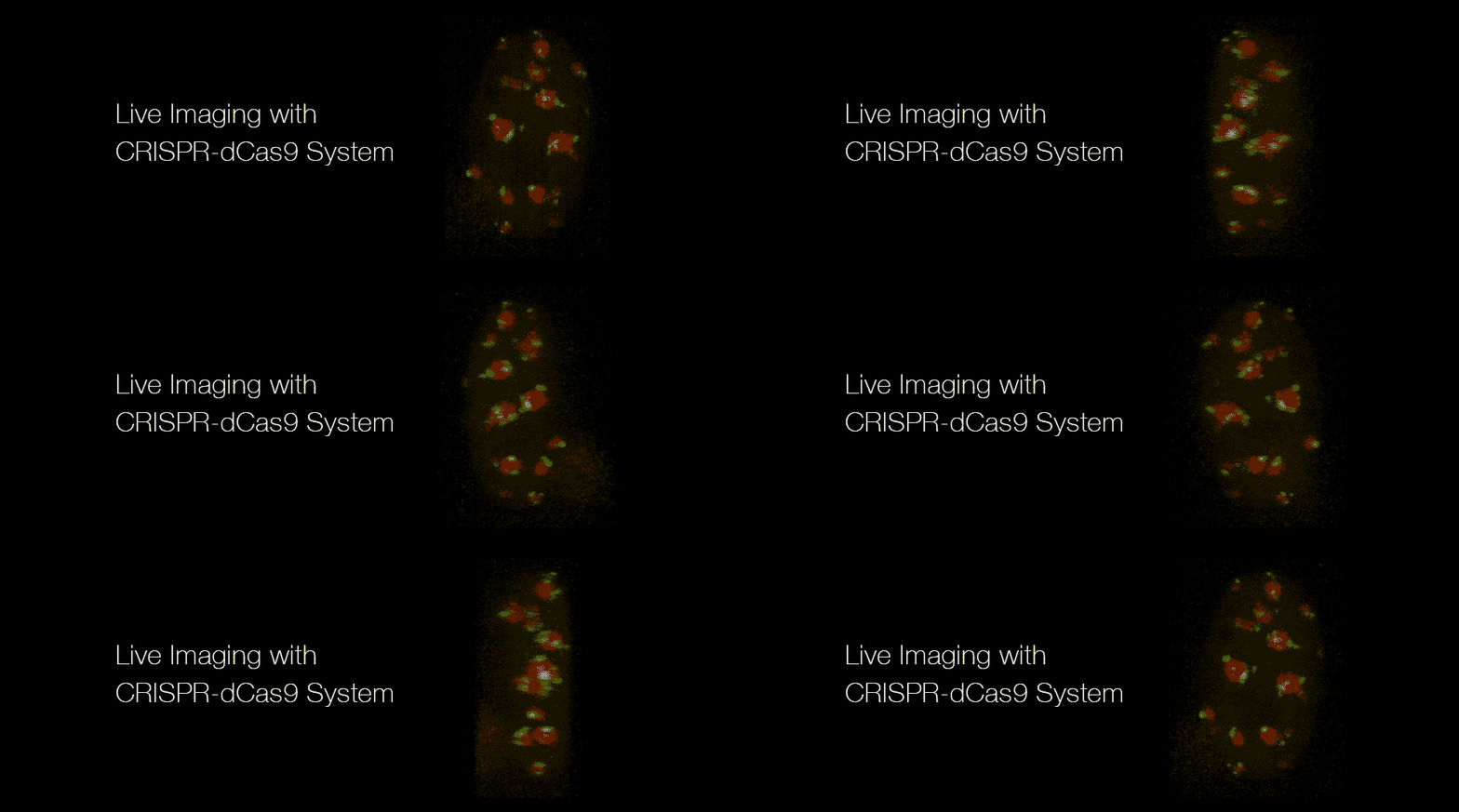
We build and apply experimental and computational approaches, including 3D DNA-FISH, dual-colour live-cell CRISPR imaging, EpiMethylTag, MethNet, single-molecule nano-NOMe phasing and computational modelling of single-cell multiome data. We use these approaches to understand transcriptional buffering, mitotic bookmarking, quiescence and differentiation, alongside chromatin reprogramming by oncogenic drivers such as NSD2 and the lasting effects of DNA damage.
In HER2-positive breast cancer, we test drug combinations to prevent the emergence of drug-tolerant persister (DTP) cells and define the mechanisms underlying their effects on chromatin, gene regulation and cell survival.
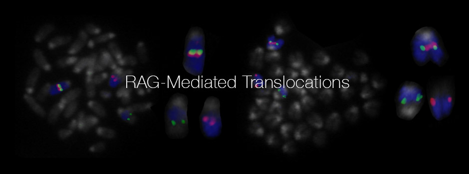
Our work helped establish how nuclear organization regulates V(D)J recombination, allelic exclusion and genome stability during lymphocyte development. We showed how locus contraction, looping, homologous pairing and nuclear positioning coordinate antigen-receptor rearrangement, including communication between alleles that helps restrict recombination to one allele at a time. We also identified how ATM and the RAG2 C-terminus control RAG cleavage, limiting simultaneous DNA breaks at different antigen-receptor loci and protecting against chromosomal translocations.
Our studies of immunoglobulin class-switch recombination (CSR) linked chromatin architecture and long-range regulatory interactions to transcription and the order of DNA break formation. We also showed how nuclear proximity contributes to AID-mediated chromosomal translocations, connecting antibody diversification with genome instability and cancer.
These discoveries laid the foundation for our broader work on genome organization and gene regulation. Today, we investigate how CTCF, CTCFL, cohesin, condensin II and histone H1 variants shape chromatin and regulate and buffer transcription. We connect these mechanisms to differentiation, quiescence and mitotic bookmarking, and investigate their disruption in cancer and developmental disorders.
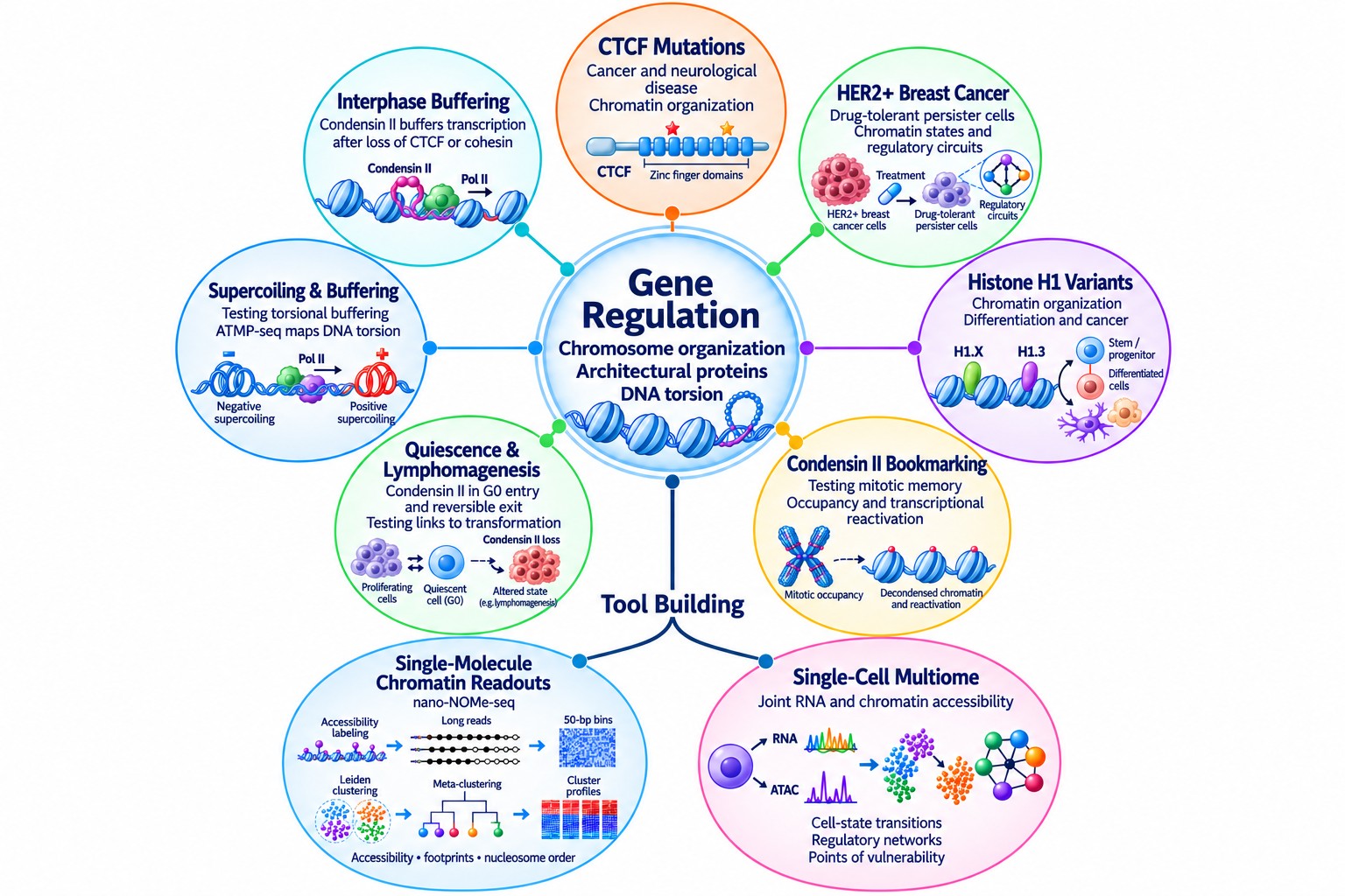
New projects ask how chromosome organization, architectural proteins and DNA torsion work together to regulate genes, in development and in disease.
How CTCF, its paralog CTCFL, cohesin and transcription factors set boundaries between chromatin domains, and what makes a boundary strong or weak in a given cell type.
We defined where the two proteins overlap and where they differ in binding, insulation and promoter control. This explains when CTCFL can substitute for CTCF, and when it works against it.
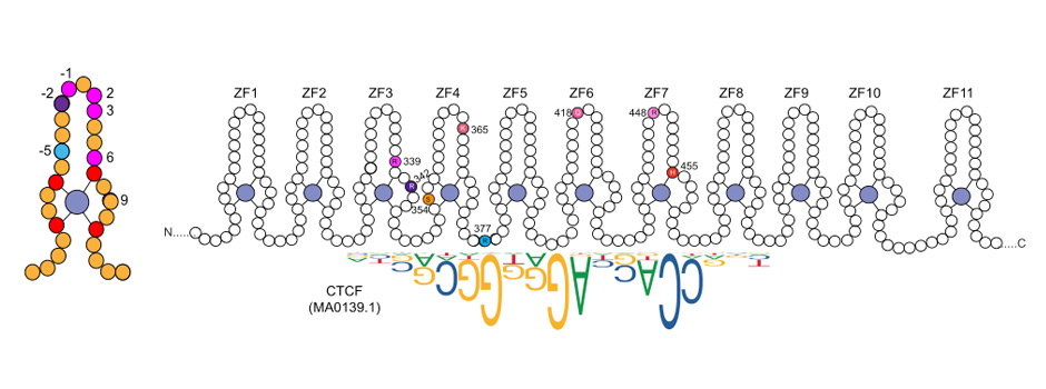
A domain-by-domain series of CTCF mutants separates how strongly CTCF binds DNA from how well it organizes chromatin. The result is a map from genotype to insulation and folding, with constraints on crosstalk between nucleosomes and transcription factors.
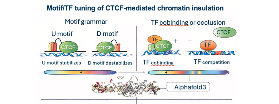
We investigate how local DNA sequence and cell-type-specific transcription factors influence CTCF and cohesin occupancy, nucleosome organization and genome folding.
Our work shows that sequences flanking the CTCF motif and nearby transcription factors can enhance or reduce CTCF binding, depending on context. Stronger CTCF occupancy does not necessarily mean stronger insulation: increased binding can coexist with disrupted nucleosome phasing and weaker boundaries. Across the sites we examined, nucleosome phasing tracks insulation strength more closely than CTCF ChIP-seq peak height.
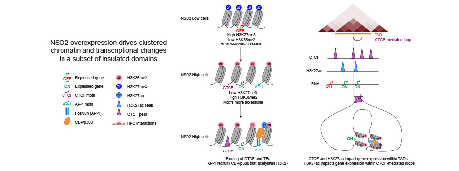
What happens to insulated domains when cancer drivers or DNA damage disturb them, and how those changes last.
NSD2 overexpression drives clustered changes in chromatin and transcription within insulated domains, establishing a domain-level mechanism for how an oncogene acts.
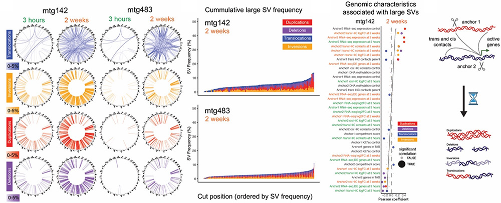
Widespread, targeted double-strand breaks cause lasting changes to chromatin and gene expression in glioblastoma cells, with implications for therapy resistance.
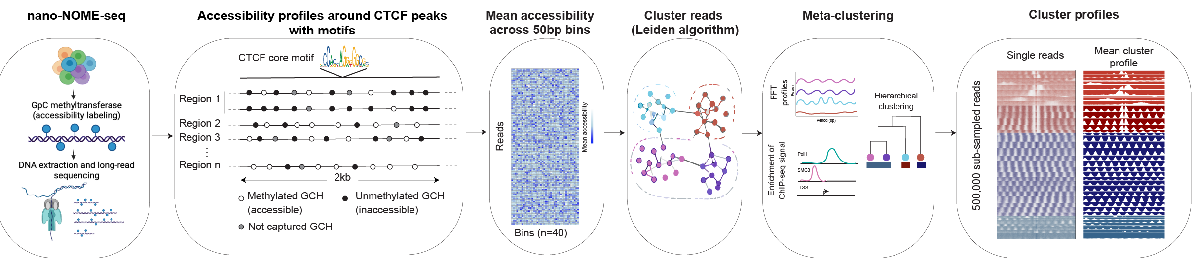
Imaging, sequencing and computational methods we have built, standardized and shared for seeing chromatin in single cells and single molecules.
Clusters single long-read molecules and stitches overlapping reads to cover long regulatory neighbourhoods. This links CTCF binding states, nucleosome occupancy and long-range interactions directly to transcription.
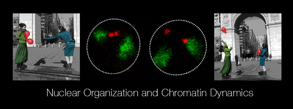
Extensive 3D DNA-FISH and immuno-FISH workflows to measure where loci sit in the nucleus, how they contract and decontract, and how higher-order architecture changes. These methods run through our papers.
CRISPR-dCas9 with sgRNA scaffolds tracks repeats and individual loci in two colours in living cells (Fu et al., Nature Communications, 2016).
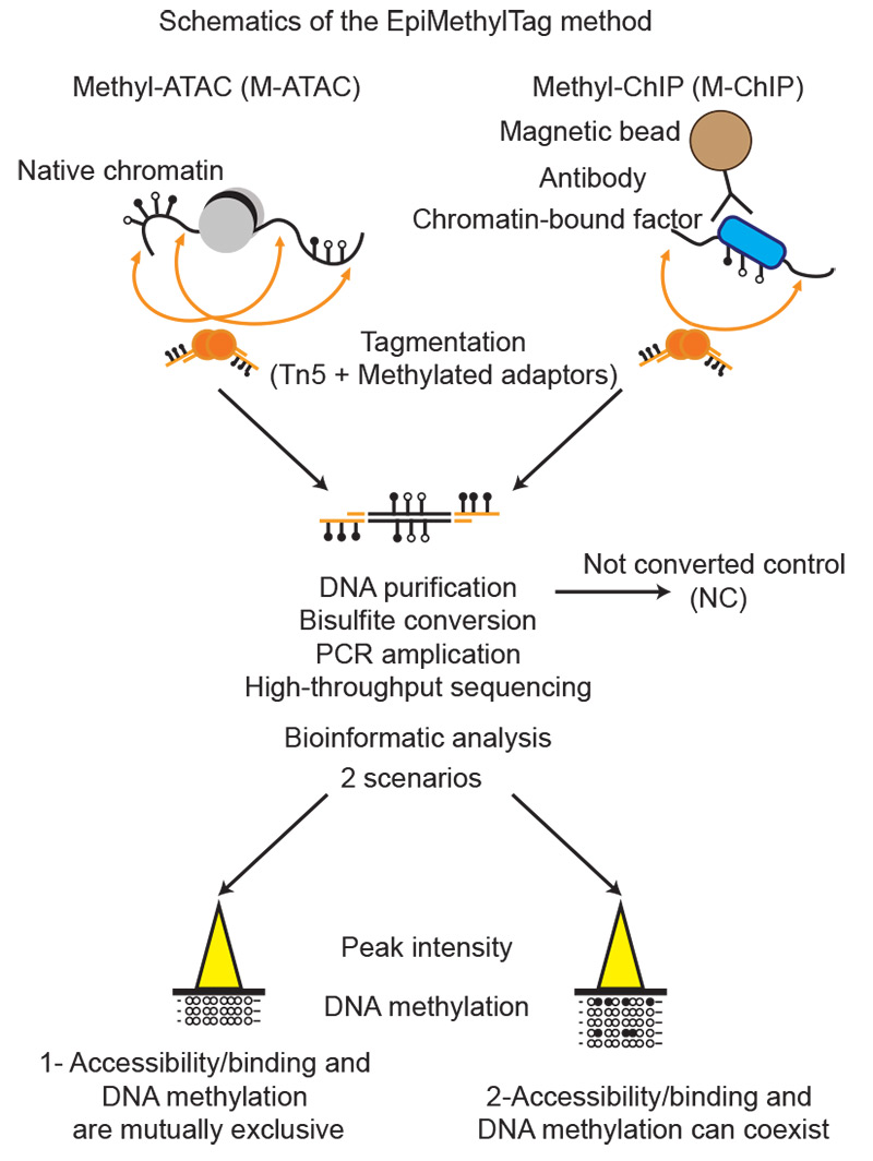
Reads ATAC-seq or ChIP-seq signal together with DNA methylation from the same material. A full protocol is available.
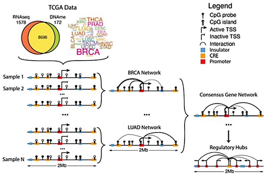
Builds methylation-anchored networks across patient cohorts to find distal regulatory hubs and the genes they control in cancer.
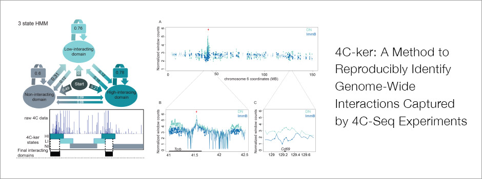
4C-ker calls 4C-seq interactions reproducibly, and a companion framework identifies simultaneous interactions within multi-loci hubs.
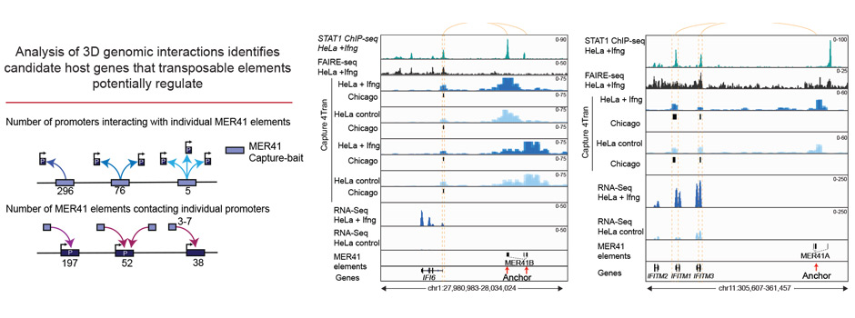
Genome-wide 3D analysis that nominates host genes likely to be regulated by transposable elements.
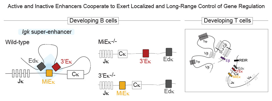
Our foundational work on how developing B and T cells assemble their antigen receptors. It set the approach that still anchors our cancer and immunity studies.
Using 3D DNA-FISH, we visualized how antigen-receptor loci fold within individual nuclei, bringing widely separated gene segments together for recombination. Our work established a role for Pax5 in Igh contraction and distal V-gene rearrangement, and showed that Tcrα and Tcrβ undergo developmentally regulated contraction through looping. These findings connected chromosome folding with access to a broad repertoire of antigen-receptor gene segments.
Papers: Fuxa et al., Genes & Development, 2004; Skok et al., Nature Immunology, 2007
We identified how changes in locus conformation and nuclear positioning contribute to allelic exclusion. At Igh, decontraction separates distant V segments from the DJ region, limiting further rearrangement, while recruitment of one allele to pericentromeric heterochromatin provides an additional layer of restriction. At Igκ, the two alleles acquire different chromatin environments before rearrangement. Together, these mechanisms help regulate access to individual alleles and support expression of a single functional receptor specificity.
Papers: Roldán et al., Nature Immunology, 2005; Goldmit et al., Nature Immunology, 2005
We showed that Igh and Igκ associate in a lineage- and developmental-stage-specific manner as B cells transition from heavy-chain to light-chain rearrangement. The 3′ Igκ enhancer is required for this association, pericentromeric recruitment and Igh decontraction. Deleting the enhancer disrupts these events and increases distal Igh rearrangement, revealing how an enhancer can regulate recombination at a locus on another chromosome.
Our work showed that nuclear positioning and enhancer activity help determine which antigen-receptor loci become available for rearrangement in developing B and T cells. We subsequently demonstrated that Igκ enhancers cooperate within a local regulatory hub in B cells. In developing T cells, the locally inactive Igκ enhancer MiEκ contacts the active Tcrβ enhancer Eβ and influences CBFβ binding and Tcrβ recombination. An enhancer can therefore regulate another locus even when its own locus is inactive.
We also linked recombination timing to developmental signals. IL-7R/STAT5 signalling coordinates Rag expression with the cell cycle. In fetal liver, reduced signalling permits earlier Igκ rearrangement, enabling a pathway that bypasses pre-BCR selection and helps generate the distinctive B-1a receptor repertoire.
Papers: Kosak et al., Science, 2002; Proudhon et al., Cell Reports, 2016; Johnson et al., Journal of Immunology, 2012; Wong et al., Nature Communications, 2019
We showed that the 3′ region of Tcrα loops outside its chromosome territory on one allele, linking transcription and increased accessibility with focal RAG binding and cleavage. ATM-dependent regulation helps restrict cleavage to one allele at a time. This work connected the spatial organisation of recombination centres with targeted DNA cleavage and genome stability.
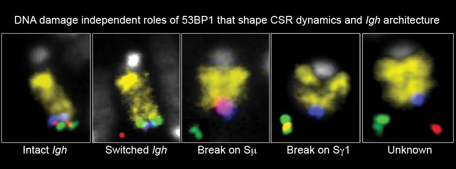
Our work linked Igh architecture to the regulation of class-switch recombination. We identified a role for 53BP1 in controlling locus conformation and the order of switch-region DNA breaks before its established function in repair. Collaborative studies showed how Mediator supports contacts between switch regions and enhancers and their transcriptional activation. We also demonstrated that proximity to Igh contributes to AID-mediated translocation partner selection, connecting nuclear organisation with the risk of oncogenic rearrangements.
Papers: Rocha et al., Molecular Cell, 2012; Thomas-Claudepierre et al., Journal of Experimental Medicine, 2016; Rocha et al., Cell Reports, 2016